
Page 15 - RETIC_Vol_4_1
P. 15
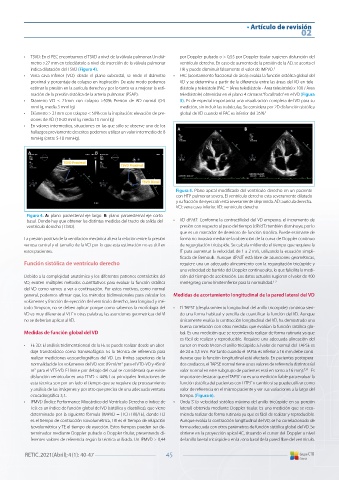
• Artículo de revisión
                                                                                                           02


          3.  Clark TJ,  Sheehan FH,  Bolson EL. Characterizing the normal heart using   the American Society of Echocardiography and the European Association
            quantitative three-dimensional echocardiography. Physiol Meas 2006; 27:   of Cardiovascular Imaging. J Am Soc Echocardiogr 2015; 28: 1-39.
            467-508.                                          10. Kjaergaard J,  Snyder EM,  Hassager C, et al. Impact of preload and af-
          4.  Rudski LG,  Lai WW, Afilalo J, et al. Guidelines for the echocardiographic   terload on global and regional right ventricular function and pressure:
            assessment of the right heart in adults: a report from the American Socie-  a quantitative echocardiography study. J Am Soc Echocardiogr 2006; 19:
            ty of Echocardiography endorsed by the European Association of Echo-  515-521.
            cardiography, a registered branch of the European Society of Cardiology,   11. Kukulski T, Hubbert L, Arnold M, et al. Normal regional right ventricular
            and the Canadian Society of Echocardiography. J Am Soc Echocardiogr   function and its change with age: a Doppler myocardial imaging study. J
            2010; 23: 685-713.                                  Am Soc Echocardiogr 2000; 13:194-204.
          5.  Kovalova S, Necas J, Cerbak R, et al. Echocardiographic volumetry of the   12. Hsiao SH,  Lin SK,  Wang WC, et al. Severe tricuspid regurgitation shows
            right ventricle. Eur J Echocardiogr. 2005; 6: 15-23.  significant impact in the relationship among peak systolic tricuspid annu-
          6.  Bommer W,  Weinert L,  Neumann A, et al. Determination of right atrial   lar velocity, tricuspid annular plane systolic excursion, and right ventricu-
            and right ventricular size by two-dimensional echocardiography. Circu-  lar ejection fraction. J Am Soc Echocardiogr. 2006; 19: 902-910.
            lation 1979; 60: 91-100.                          13. Ghio S,  Recusani F,  Klersy C, et al. Prognostic usefulness of the tricuspid
          7.  Van der Zwaan HB,  Helbing WS,  McGhie JS, et al. Clinical value of real-  annular plane systolic excursion in patients with congestive heart failure
            time three-dimensional echocardiography for right ventricular quantifi-  secondary to idiopathic or ischemic dilated cardiomyopathy. Am J Cardiol
            cation in congenital heart disease: validation with cardiac magnetic reso-  2000; 85: 837-842.
            nance imaging. J Am Soc Echocardiogr 2010; 23: 134-140.  14.  Vivo R,  Cordero-Reyes A, Qamar U, et al. Increased right-to-left ventricular
          8.  Weidemann F,  Eyskens B,  Mertens L, et al. Quantification of regional left   diameter ratio is a strong predictor of right ventricular failure after left
            and right ventricular function by ultrasonic strain rate and strain indexes   ventricular assist device. J Heart Lung Transplant 2013; 32:792-799.
            after surgical repair of tetralogy of Fallot. Am J Cardiol. 2002; 90: 133-138.   15.  Goldraich L,  Kawajiri H,  Foroutan F, et al.: Tricuspid valve annular dilata-
          9.  Lang RM,  Badano LP,  Mor-Avi F, et al. Recommendations for cardiac   tion as a predictor of right ventricular failure after implantation of a left
            chamber quantification by echocardiography in adults: an update from   ventricular assist device. J Cardiol Surg. 2016; 31:110-116.


          RETIC. 2021(Abril); 4 (1):  40-47                47